本會期刊
台灣急診醫學通訊 
第四卷第二期
刊登日期:2021/04/29
Taiwan Emergency Medicine Bulletin 4(2) : e2021040202回上頁
點閱次數:2182
PDF下載次數:3
頸動脈超音波於急診之應用
陳麒心  范程羿 宋之維 黃沛銓 范程羿 宋之維 黃沛銓 臺大醫院新竹分院 急診醫學部
焦點式超音波(Point-of-care ultrasound, POCUS)現今已是急診醫師不可或缺的技能。焦點式超音波涵蓋範圍極廣,在腹部、胸腔、心臟、血管、骨骼及軟組織等急症的診斷及侵入性治療的導引皆扮演重要的角色1。最近隨著急診焦點式超音波及其相關研究的蓬勃發展,超音波的應用更導入了急救復甦的流程,提供了心跳停止原因之鑑別、檢視氣管內管的置放、甚至檢查心臟是否有搏動等的協助2。然而,在眾多焦點式超音波的應用中,針對神經系統疾病檢查的頸動脈超音波(carotid ultrasound)及顱內血管超音波(Transcranial ultrasound)則相對較少被討論及使用。其中,頸動脈超音波因其較易學、執行也相對快速等可近性,以及許多潛在的應用,有越來越多研究探討在急診使用焦點式頸動脈超音波的可行性3,4。以下就頸動脈超音波之幾個主要用途及潛在應用加以介紹 1. 檢查頸動脈粥狀樣化及狹窄
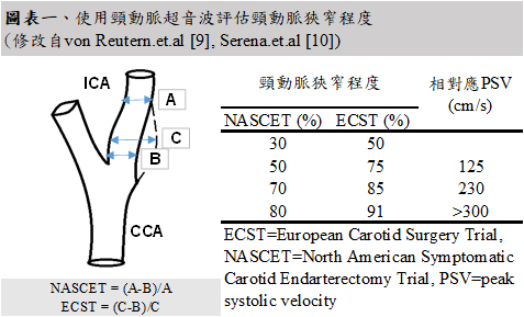
偵測頸動脈狹窄程度及斑塊為頸動脈超音波最主要的用途。過去研究發現,頸動脈粥狀硬化造成的頸動脈斑塊與頸動脈狹窄佔缺血性中風病因的百分之十五到二十5,也是暫時性腦缺血後再次中風的重要危險因子6。針對有症狀且狹窄程度大於70%的病人,介入性之治療如頸動脈內膜切除術、支架置放等亦被證實有其必要性,然而需在腦缺血事件發生的兩周內治療才能最大化其好處7,8。而頸動脈超音波相較於過去視為診斷標準的電腦斷層血管攝影具有能快速操作、不須施打顯影劑以及無輻射暴露等優點。頸動脈超音波的所被測量的參數眾多,在狹窄程度的測量,可在B-mode下使用北美(NASCET)或歐洲(ECST)的測量方法進行量測,或使用都普勒超音波(doppler ultrasound)測量內頸動脈尖峰收縮期流速進行推估(如圖表一)9, 10。除了狹窄程度,頸動脈斑塊的偵測亦是重要的檢查項目,而針對斑塊及其特徵的描述,常見的包括同質/異質性、表面規則度、超音波回聲度、斑塊潰瘍等11。然而要進行以上完整的頸動脈都普勒超音波評估,仍需不少時間,且需長時間的訓練或進階的超音波儀器方能執行。故如何能在暫時性腦缺血及缺血性腦中風的急診病人身上快速施行焦點式的頸動脈超音波,進行風險評估及導引治療仍為一大課題。Robert Suttie等人於2020年所發表的研究,針對於急診暫時性腦缺血及缺血性腦中風的病人進行頸動脈超音波掃描3。此研究中以肉眼評估(eyeball evaluation)判斷其是否為高程度(>50%)之頸動脈狹窄,並與電腦斷層血管攝影之結果比較,發現能達到敏感度70%、特異度86.7%之診斷效力,並平均能在6分鐘左右內完成3。 在疑似或確診暫時性腦缺血及缺血性腦中風的病人身上快速施行焦點式的頸動脈超音波亦有機會能診斷頸動脈剝離甚至間接發現主動脈剝離,並減少診斷錯誤或誤判為缺血性中風而施打血栓溶解劑可能造成的潛在致命性併發症。總歸以上之好處與應用性,頸動脈超音波可做為疑似或確診暫時性腦缺血及缺血性腦中風的病人的焦點式超音波檢查項目,而日後研究更可針對此設計一符合急診焦點式超音波使用之診斷流程,以期達到能在不延遲stroke code的診斷治療下快速操作,又能達到更好的診斷效力。 
2. 輸液治療反應(fluid responsiveness)之評估
輸液治療反應的評估一直是急重症領域相當火熱的話題,而超音波亦是其中一項很重要的評估工具。除可從基本的下腔靜脈的直徑與呼吸時的塌陷程度,至進階的心臟超音波如左心室開口-主動脈流速對時間積分(velocity time integral, VTI)等,頸動脈超音波亦是其中之一12。被動抬腿測試(passive leg raising, PLR)後使用頸動脈超音波量測頸動脈矯正收縮期流動時間(carotid corrected systolic flow time, ccFT)被證實能提供輸液治療反應的依據13。研究亦發現利用短短5分鐘的教學影片,即能讓急診住院醫師有良好的操作能力,亦更證明了這項技術的可近性4。因此,日後急診醫師使用焦點式超音波進行輸液治療反應的評估時,亦可加入頸動脈超音波的掃描做為輔助。 3. 於復甦急救之應用
頸動脈超音波於復甦急救之應用與研究包括急救中檢查脈搏(check pulse)與藉由頸動脈血流評估壓胸品質14, 15。然而頸動脈超音波於復甦急救之應用之研究仍較少且分歧,應是未來急診醫師可努力之方向。 總而言之,因頸動脈超音波具廣泛的用途與臨床價值及可近性,應是急診醫師進行焦點式超音波掃描與學習的一重要技能。如何快速且準確地進行標準化頸動脈超音波掃描更是日後研究需努力的方向。
參考文獻
1. Moore CL, Copel JA. Point-of-care ultrasonography. N Engl J Med. 2011 Feb 24;364(8):749-57.
2. Lien WC, Hsu SH, Chong KM, et al. US-CAB protocol for ultrasonographic evaluation during cardiopulmonary resuscitation: Validation and potential impact. Resuscitation. 2018 Jun;127:125-131.
3. Suttie R, Woo MY, Park L, Nemnom MJ, Stotts G, Perry JJ. Can Emergency Physicians Perform Carotid Artery Point-of-Care Ultrasound to Detect Stenosis in Patients with TIA and Stroke? A Pilot Study. West J Emerg Med 2020;21(3):626-632.
4. Stolz LA, Mosier JM, Gross AM, Douglas MJ, Blaivas M, Adhikari S. Can emergency physicians perform common carotid Doppler flow measurements to assess volume responsiveness? West J Emerg Med. 2015 Mar;16(2):255-9.
5. Joh JH, Cho S. Cardiovascular risk of carotid atherosclerosis: global consensus beyond societal guidelines. Lancet Glob Health. 2020 May;8(5):e625-e626.
6. Giles MF, Rothwell PM. Risk of stroke early after transient ischaemic attack: a systematic review and meta-analysis. Lancet Neurol. 2007 Dec;6(12):1063-72.
7. North American Symptomatic Carotid Endarterectomy Trial Collaborators, Barnett HJM, Taylor DW, et al. Beneficial effect of carotid endarterectomy in symptomatic patients with high-grade carotid stenosis. N Engl J Med. 1991 Aug 15;325(7):445-53.
8. Gladstone DJ, Oh J, Fang J, et al. Urgency of carotid endarterectomy for secondary stroke prevention: results from the Registry of the Canadian Stroke Network. Stroke. 2009 Aug;40(8):2776-82.
9. von Reutern GM, Goertler MW, Bornstein NM, et al. Grading carotid stenosis using ultrasonic methods. Stroke. 2012 Mar;43(3):916-21.
10. Serena J, Irimia P, Calleja S, Blanco M, Vivancos J, Ayo-Martín O; Representación de la Sociedad Española de Neurosonología (SONES). Cuantificación ultrasonográfica de la estenosis carotídea: recomendaciones de la Sociedad Española de Neurosonología [Ultrasound measurement of carotid stenosis: recommendations from the Spanish Society of Neurosonology]. Neurologia. 2013 Sep;28(7):435-42.
11. Brinjikji W, Rabinstein AA, Lanzino G, Murad MH, Williamson EE, DeMarco JK, Huston J 3rd. Ultrasound Characteristics of Symptomatic Carotid Plaques: A Systematic Review and Meta-Analysis. Cerebrovasc Dis. 2015;40(3-4):165-74.
12. Pourmand A, Pyle M, Yamane D, Sumon K, Frasure SE. The utility of point-of-care ultrasound in the assessment of volume status in acute and critically ill patients. World J Emerg Med. 2019;10(4):232-238.
13. Barjaktarevic I, Toppen WE, Hu S, et al. Ultrasound Assessment of the Change in Carotid Corrected Flow Time in Fluid Responsiveness in Undifferentiated Shock. Crit Care Med. 2018 Nov;46(11):e1040-e1046.
14. Sanchez S, Miller M, Asha S. Assessing the validity of two-dimensional carotid ultrasound to detect the presence and absence of a pulse. Resuscitation. 2020 Dec;157:67-73.
15. Yilmaz G, Silcan M, Serin S, Caglar B, Erarslan Ö, Parlak İ. A comparison of carotid doppler ultrasonography and capnography in evaluating the efficacy of CPR. Am J Emerg Med. 2018 Sep;36(9):1545-1549.
回上頁
|
 登入/註冊
登入/註冊

